
Dog Skeleton Chart, an art print by Roselyne Lougnon INPRNT
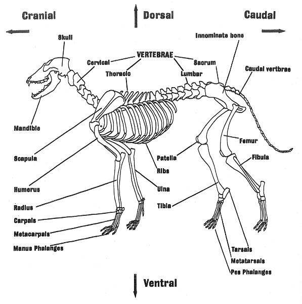
Anatomic Planes. The main planes of motion for dogs are as follows (see Figure 5-1): • The sagittal plane divides the dog into right and left portions. If this plane were in the midline of the body, this is the median plane or median sagittal plane. • The dorsal plane divides the dog into ventral and dorsal portions.

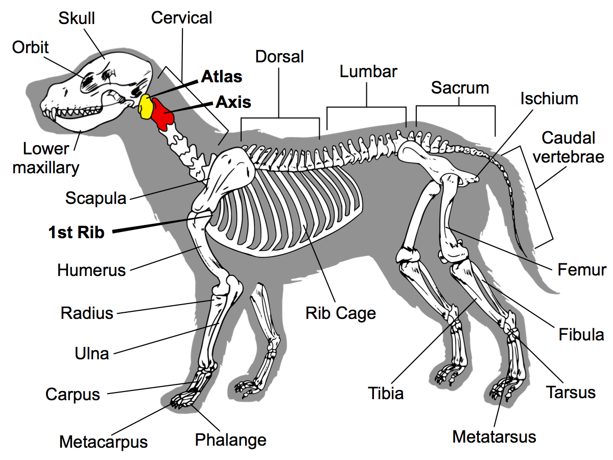
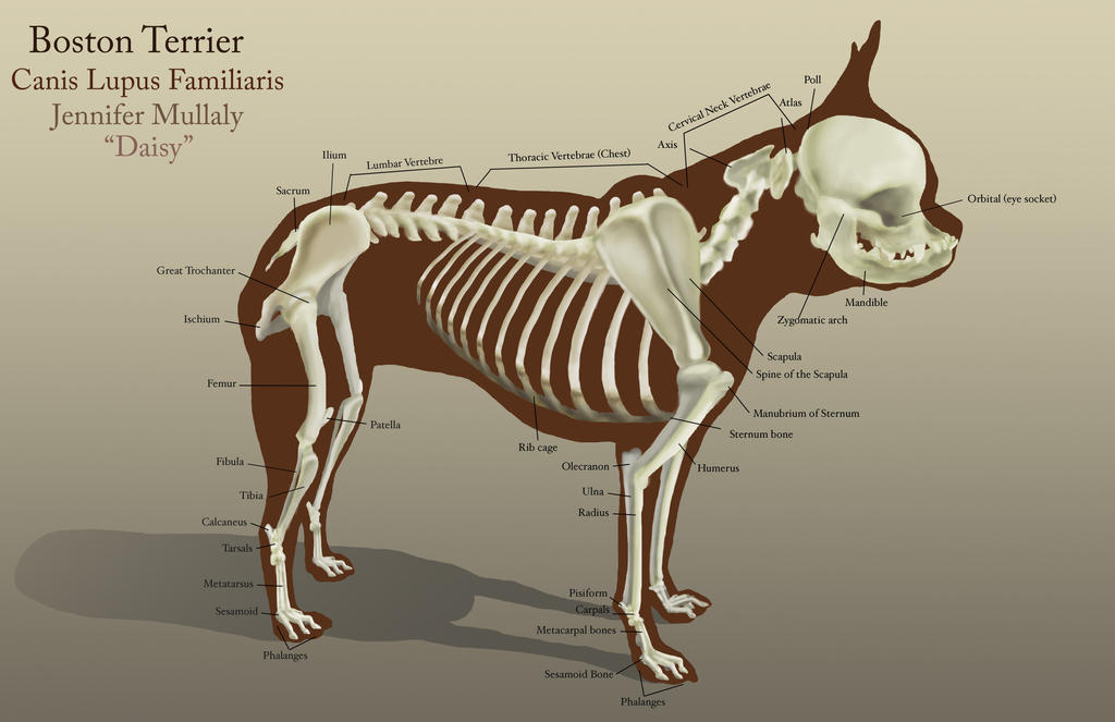
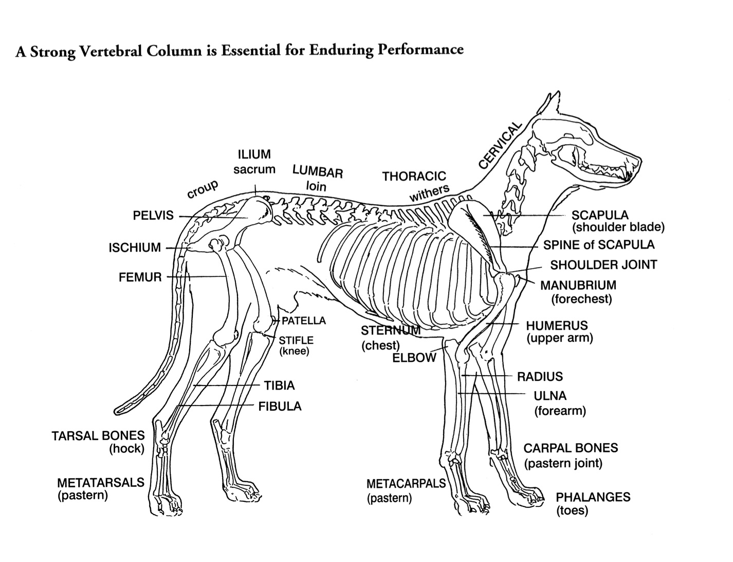
Dog skeleton with major bone elements labeled (Davis, 1987, p. 54
Whereas giant breeds can take between 18 months and 2 years for their growth plates to fuse. Speaking of skeletons, a dog has 320 bones in their body (depending on the length of their tail) and around 700 muscles. Muscles attach to bones via tendons. Depending on the breed of dog, they will have different types of muscle fibers.

PetMassage Chart 3 Skeleton of the Dog · PetMassage™ Training and
2021 Ultimate Guide to Dog Anatomy. As the pace of veterinary advancement accelerates, even the most experienced veterinary teams are challenged to keep up with all the changes that impact their practice.. potential outcomes and prognoses and tools e.g. anatomical diagrams, clinical formulas and programs and home care videos; Enhancing the.
A Visual Guide To Dog Anatomy Muscle Organ Skeletal
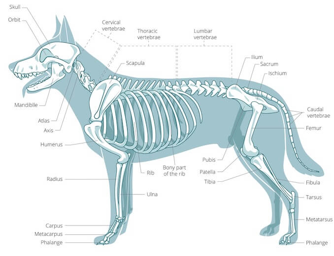
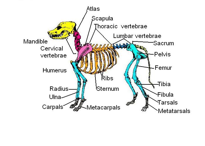
Here, I will provide a dog skeleton labeled diagram and the different parts of a dog diagram. In the dog skeleton labeled diagram, I tried to show you all the bones from the body. This might help you understand the different regions of the body so quickly. I would like to show different external features of a dog again here in a labeled picture.
Dog Skeleton Anatomy by TheDragonofDoom on DeviantArt
Animal Anatomy (Veterinary Diagrams) Dog Skeletal Anatomy. High Resolution PDF for Printing. Click Here. Link to More Information About This Animal. Click Here. Citing Research References.. Amsel, Sheri. "Dog Skeletal Anatomy" Exploring Nature Educational Resource ©2005-2024. January 10, 2024
Anatamation where Anatomy meets Animation Dog anatomy
1. Dog Anatomy: A Pictorial Approach. Dog Anatomy: A Pictorial Approachby Peter C. Goody offers clear and precise illustrations of the skeletal-muscular system of canines. Each diagram is meticulously labeled with little additional text since the book truly takes a pictorial approach to the topic.
Home Study “Canine Musculoskeletal Unwinding” Watch Instantly Video
Download scientific diagram | Dog skeleton with major bone elements labeled (Davis, 1987, p. 54; Reitz & Wing, 2008, p. 364). from publication: Zooarchaeological analysis of bones from.

Easy Diagram Of Backbone Binding of phosphate backbone of DNA to the
Components of the Musculoskeletal System in Dogs. Bones provide rigid structure to the body and shield internal organs from damage. They also house bone marrow, where blood cells are formed, and they maintain the body's reservoirs of calcium and phosphorus. Old bone tissue is constantly replaced with new bone tissue in a process called.
Skeleton Worksheet Answers WikiEducator
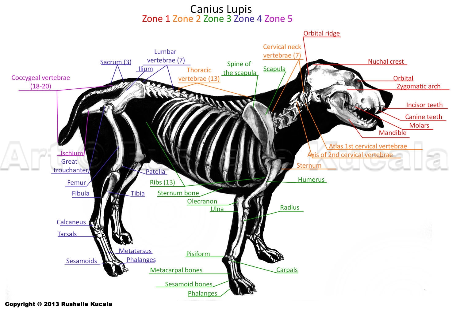
ISSN 2534-5087. This veterinary anatomy module of the dog contains 218 illustrations dedicated to the canine osteology anatomy. Here are presented scientific illustrations of the canine skeleton, with the main dog's bones and its structures displayed from different anatomical standard views (cranial, caudal, lateral, medial, dorsal, palmar..).
Dog Skeleton by peanutbutterjenny on DeviantArt
Labeled anatomy of the head and skull of the dog on CT imaging (bones of cranium, brain, face, paranasal sinus, muscles of head) This module of vet-Anatomy presents an atlas of the anatomy of the head of the dog on a CT. Images are available in 3 different planes (transverse, sagittal and dorsal), with two kind of contrast (bone and soft tissues).
Types and Parts of Bones Reading Ancient Animal Remains
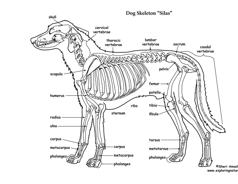
Your dog's skeletal system provides the body's fram ework and structure as well as protects many of its organs. Did you know there are over 30 0 bones in a dog? Can you correctly identify some of the bones on the diagram below? Use the bones list in the shaded box to match the bones in the illustrati on. Write the correct body part number.

Dog Vertebral Column Anatomy ANATOMY STRUCTURE
A dog's skeleton is made up of many different bones, which provide structure and support for their body. Dogs have over 300 bones in their body, which is more than humans who have around 206 bones. Their skeleton includes their skull, spine, ribcage and limbs. Dogs have four legs that are designed to help them move quickly and efficiently.

Anatomy Of Dog Skeleton With Labeled Inner Bone Scheme Vector
Dog skeleton. As with any vertebrate animal, the skeleton of a dog has the function of supporting the body for movement and protecting its internal organs. We can divide the canine skeleton into three main sections: Axial skeleton: skull, spine, ribs and sternum bones. Appendicular skeleton: bones of the extremities.

Skeletal System Review
Anatomy atlas of the canine general anatomy: fully labeled illustrations and diagrams of the dog (skeleton, bones, muscles, joints, viscera, respiratory system, cardiovascular system). Positional and directional terms, general terminology and anatomical orientation are also illustrated.
Dog Skeletal Anatomy
Here, in the dog skeleton labeled diagram, I tried to show you the different segments of the forelimb, hindlimb with their bones. Again, I tried to show you all the bones from the vertebrae column of a dog skeleton. In addition, in the diagram, you will find a few identified skull bones. The sternum and the ribs are also identified in the dog.
Helen King on Structure Evaluation Susan Garrett's Dog Training Blog
Dog anatomy comprises the anatomical studies of the visible parts of the body of a domestic dog.Details of structures vary tremendously from breed to breed, more than in any other animal species, wild or domesticated, as dogs are highly variable in height and weight. The smallest known adult dog was a Yorkshire Terrier that stood only 6.3 cm (2.5 in) at the shoulder, 9.5 cm (3.7 in) in length.